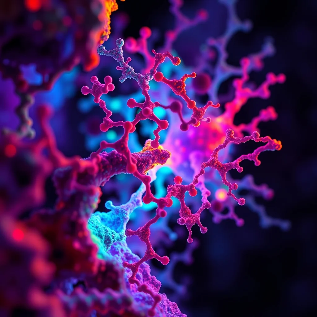
electron microscope enlargement of a cascading protein - to - protein kinase interactions, causing cancer cells division, volumetric complementary colors, refracted kodachromatic internal lighting, photorealistic, 8 k
electron microscope enlargement of a cascading protein - to - protein kinase interactions, causing cancer cells division, volumetric complementary colors, refracted kodachromatic internal lighting, photorealistic, 8 k
electron microscope enlargement of a chilopoda section, surrounded by acicular crystals, volumetric complementary colors, refracted kodachromatic internal lighting
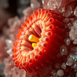
electron microscope enlargement of a chilopoda section, surrounded by acicular crystals, refracted kodachromatic internal lighting, complementary colors
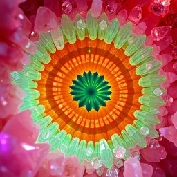
electron microscope enlargement of a chilopoda section, surrounded by acicular crystals, complementary colors, refracted kodachromatic internal lighting, god rays
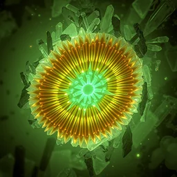
electron microscope enlargement of a chilopoda section, surrounded by acicular crystals, refracted kodachromatic internal lighting, various autochromatic lumiere photogram
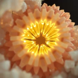
electron microscope enlargement of a chilopoda section, surrounded by acicular crystals, refracted internal light, kodachromatic
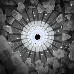
electron microscope enlargement of a chilopoda section, surrounded by acicular crystals, refracted kodachromatic internal lighting

a protein, a protein, a protein
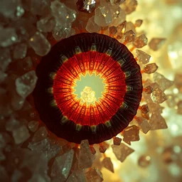
electron microscope enlargement of a chilopoda section, surrounded by acicular crystals, refracted internal light, autochrome lumiere
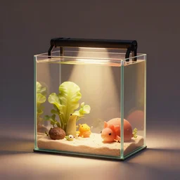
Miniature aquarium, by Justin Gerard and Haeckel. Artstation, volumetric light, ray of light, warm colors
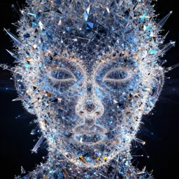
fractal - patterned face made of crystal, kodachromatic, dynamic lighting, 8 k
fractal - patterned face made of crystal, kodachromatic, dynamic lighting, volumetric golden lighting background, 8 k
high quality render of bacteria infecting healthy cells biology virus microscope

complementary colors
a microscopy image showing a cell with a normal nucleus and one cell with an abnormal stained nucleus, confocal miscroscopy, paper, cell journal, cell biology, nature journal, science
electron microscope enlargement of a chilopoda section, surrounded by acicular crystals, refracted internal light, rayograph photography
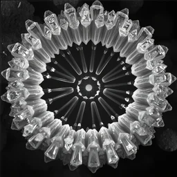
electron microscope enlargement of a chilopoda section, surrounded by acicular crystals, refracted internal light
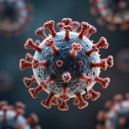
Electron microscope image of coronavirus

Stephen Hawking at the bottom of the ocean volumetric lighting, HDR, polished, micro details, ray tracing, 8k
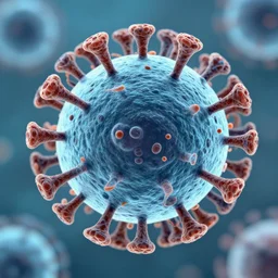
electron microscope image of a cooties virus

white blood cell, microscopy
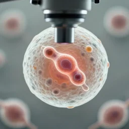
a singular cell undergoing cell division as seen through a microscope
a sideways cone with a beam of light being emitted from the bottom of the cone a raytraced image by pixar, featured on dribble, new objectivity, volumetric lighting
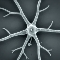
neurons and dendrites seen through scanning electron microscope