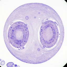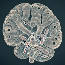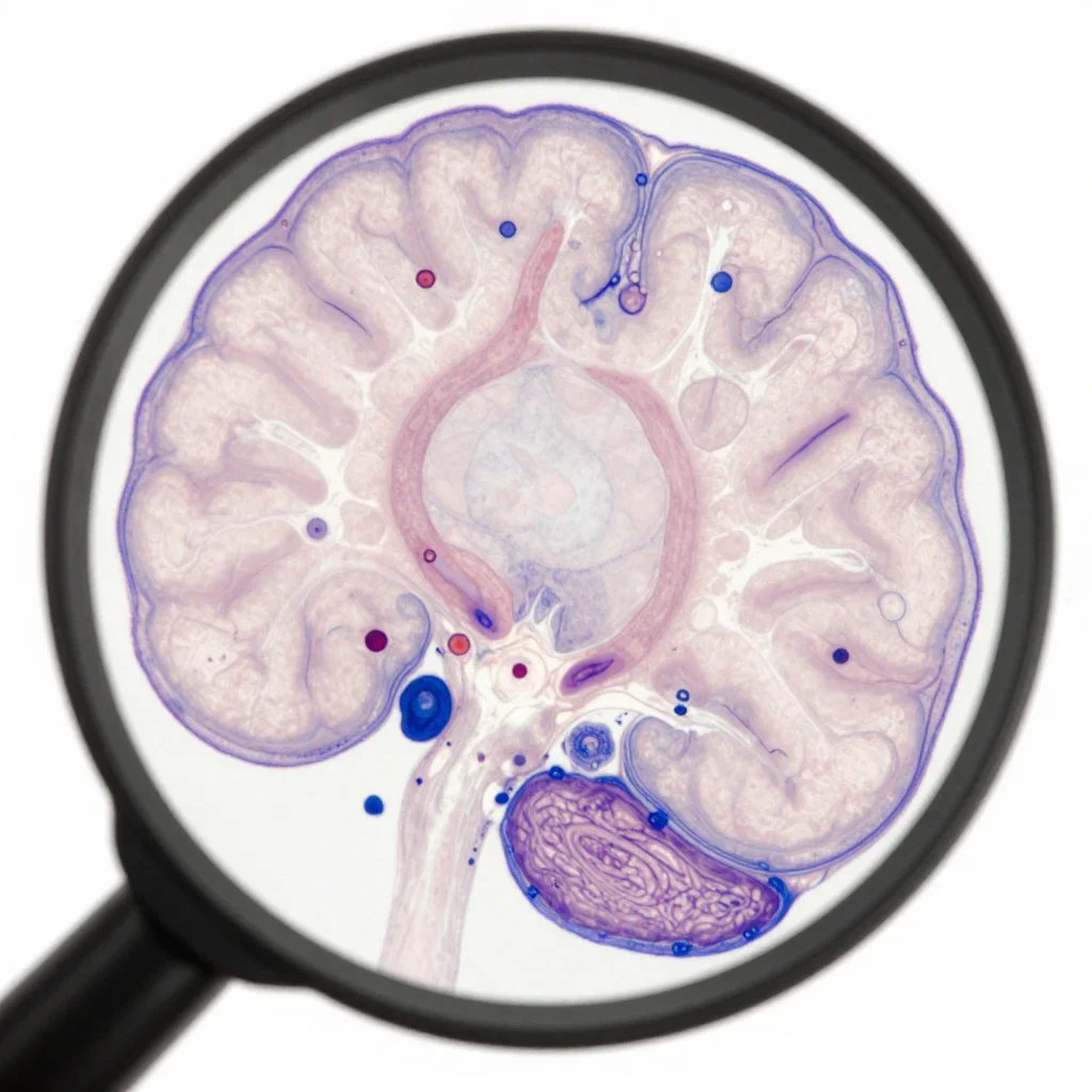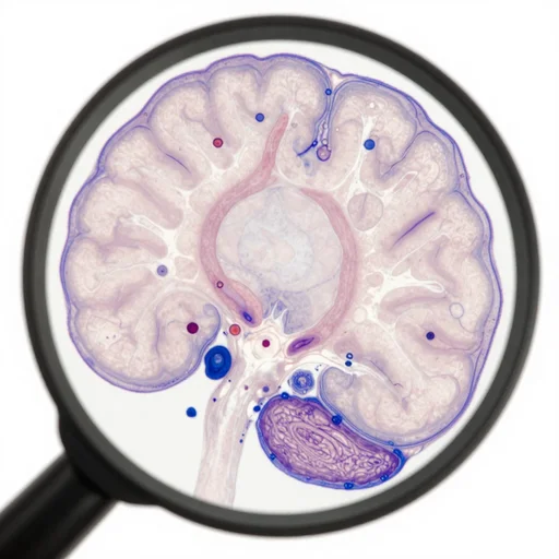
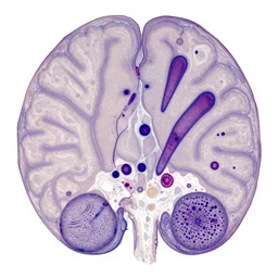
photo taken with a light microscope showing spongiform degeneration in the cerebral cortex of a patient who had died of creutzfeldt - jakob disease ( cjd ). the tissue section was embedded in paraffin - wax, sectioned at a thickness of approximately 5 - 1 0 microns, and stained with hematoxylin and eosin ( h & e ). several large, pyramid - shaped neurons can be seen, along with the nuclei of glial cells.
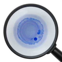
magnified 1 0 0 x, and stained with h & e ( hematoxylin and eosin ) staining ...
🎨 Related images (24)
magnified 1 0 0 x, and stained with h & e ( hematoxylin and eosin ) staining technique
hematoxylin and eosin stained breast cells under microscope, pathology image,
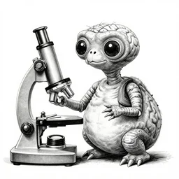
a black and white ink pen illustration of e. t. the extraterrestrial ( 1 9 8 2 ) using a microscope and holding a small brain in in his hand, highly detailed
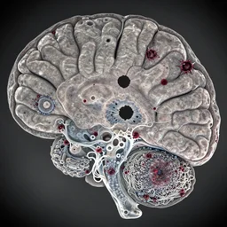
light photomicrograph of brain tissue 100X magnification showing perforation of brain matter, neural decay, synaptic patters, creepy bloody spots, holes in the brain, gloomy, eerie, swiss cheese pattern, synaptic spindles aggregations amalgamations
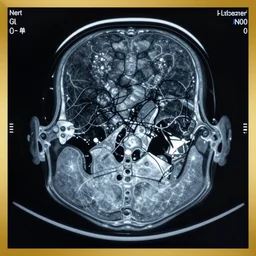
CT Scan of Alzheimer patient revealing the true cause of the disease
light photomicrograph of brain tissue 100X Magnification, synaptic patterns of nerve cells with holes in the membranes, swiss cheese pattern, synaptic spindles of neurons, many spindles of aggregations, amalgamations, entanglements, neural decay
a still photo from e. t. the extraterrestrial ( 1 9 8 2 ), e. t. is sitting at a table in a laboratory with a microscope, he is holding a little animal brain, black and white ink pen illustration, highly detailed, hyper realistic
hyper realistic portrait of professor hubert farnsworth under studio lighting | detailed, hd, cgi, realistic, photorealistic, subsurface scattering, volumetric lighting, global illumination, microdisplacement, micro detail
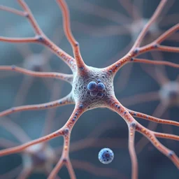
tilt shift, cells, neurons, membranes, ultra detailed
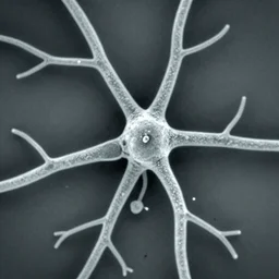
neurons and dendrites seen through scanning electron microscope
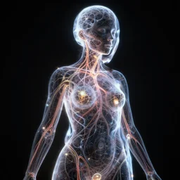
fractal mesh of a female form shaped with dark and bright neurons and veins, translucent, surface illumination, see - through, portrait, dramatic, cinematographic lighting, hyper detailed, ultra realistic, glow in the dark
a still photo from e. t. the extraterrestrial ( 1 9 8 2 ), e. t. is sitting at a table in a laboratory near a black school microscope, he is holding a broken piece of glowing rock, black and white ink pen illustration, extremely detailed, hyper realistic
brain of an nft user under the microscope
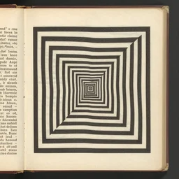
a visual illusion, 1 9 2 0 s medical textbook scan, hi - res, worn paper
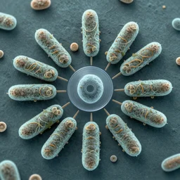
microscopy, 1 0 0 0 x magnification, electron microscope, diatoms, tardigrades, close up, centered, bacteria germs made in the style of roger dean
extremely psychedelic beautiful cyborg queen of lsd infected by night. intricate, elegant, highly detailed, extremely lifelike photorealistic digital painting, artstation. steichen, gaston bussiere, tom bagshaw, cyberpunk alphonse mucha. dark pallet, melancholy. anatomically correct in every way. sultry. sharp focus. soft light. very very lifelike
old wetplate daguerreotype portrait of the pleasure bot, explosion of data fragments, fractal, intricate, elegant, highly detailed, parallax, leica, medium format, subsurface scattering, by jheronimus bosch and greg rutkowski and louis jacques mande daguerre,
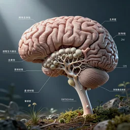
brain mold, mycelium, nervous system, cns, medical, biology, diagram overlay, high resolution, modern textbook photo
old wetplate daguerreotype portrait of the pleasure bot, explosion of data fragments, fractal, intricate, elegant, highly detailed, parallax, leica, medium format, subsurface scattering, by jheronimus bosch and greg rutkowski and louis jacques mande daguerre
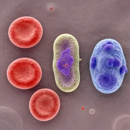
scanning electron micrograph of a red blood cell ( left ), a platelet ( center ), and a t lymphocyte ( right ) ; colorized
a still photo from e. t. the extraterrestrial ( 1 9 8 2 ), e. t. is sitting at a table in a laboratory near a vintage microscope, he is holding a broken piece of glowing rock, black and white ink pen illustration, highly detailed, hyper realistic
a singular cell undergoing cell division as seen through a microscope
hyper-detailed antique color pencil vintage medical illustration of Kaiju head cross-section, nautilus brain, ribcage, xhenomorph, with tentacles coming out of open mouth and exposed jaw bone, spinal column, cerebral corpus callosum, interventricular foramen, symmetrical, on weathered parchment paper