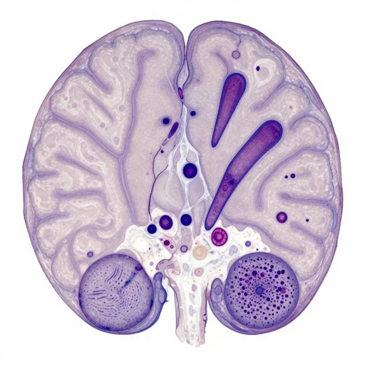
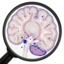
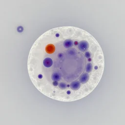
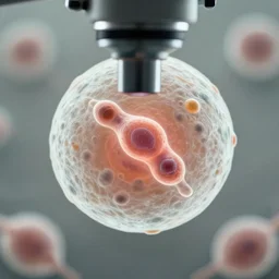
magnified 1 0 0 x, and stained with h & e ( hematoxylin and eosin ) staining technique, this light photomicrograph of brain tissue reveals the presence of prominent spongiotic changes in the cortex, and loss of neurons in a case of variant creutzfeldt - jakob disease ( vcjd ).
photo taken with a light microscope showing spongiform degeneration in the ce...
photo taken with a light microscope showing spongiform degeneration in the cerebral cortex of a patient who had died of creutzfeldt - jakob disease ( cjd ). the tissue section was embedded in paraffin - wax, sectioned at a thickness of approximately 5 - 1 0 microns, and stained with hematoxylin and eosin ( h & e ). several large, pyramid - shaped neurons can be seen, along with the nuclei of glial cells.
Details
Tags