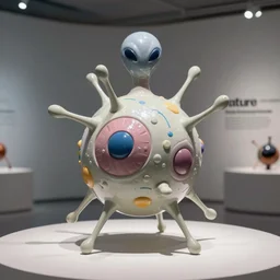
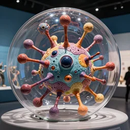
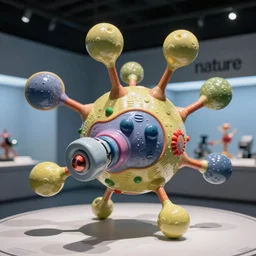
tunneling electron microscope image of broken membrane, broken leaking cell wall, liquid oxygen migrating across the cell membrane of a mitochondria oblong organelle. liquid dots penetrating crossing cell membrane edge. dividing crossing over eukaryote membrane. false color. mit technology review. cell journal. nobel prize winning. ultra detailed 8 k tiff
tunneling electron microscope image of a broken cell eukaryote lysed. false c...
🎨 Related images (24)
tunneling electron microscope image of broken membrane, broken leaking cell wall, liquid oxygen migrating across the cell membrane of a mitochondria oblong organelle. liquid stream penetrating crossing cell membrane edge. dividing crossing over eukaryote membrane. false color. mit technology review. cell science nature. nobel prize winning. ultra detailed 8 k tiff
tunneling electron microscope image of leaking cell membrane, broken leaking cell wall, liquid oxygen migrating across cell membrane of a mitochondria oblong organelle. liquid stream penetrating crossing cell membrane edge. dividing crossing over eukaryote membrane. false color. mit technology review. cell science nature. nobel prize winning. ultra detailed 8 k tiff
tunneling electron microscope image of broken membrane, broken leaking cell wall, liquid oxygen migrating across the cell membrane of a mitochondria oblong organelle. liquid dots penetrating crossing cell membrane edge. dividing crossing over eukaryote membrane. false color. mit technology review. cell science nature. nobel prize winning. ultra detailed 8 k tiff
tunneling electron microscope image of a cell quartered by an arrow. false color. mit technology review. nature journal. nobel prize winning. ultra detailed 8 k tiff
tunneling electron microscope image of a cell pierced by an arrow. line penetrating cell. false color. mit technology review. nature journal. nobel prize winning. ultra detailed 8 k tiff
tunneling electron microscope image of a mitochondria organelle. false color. mit technology review. nature journal cover. nobel prize winning. ultra detailed 8 k tiff
tunneling electron microscope image of a mitochondria organelle leaking oxygen. false color. mit technology review. nature journal. nobel prize winning. ultra detailed 8 k tiff
tunneling electron microscope image of a eukaryote pierced by a line of atoms. line penetrating cell. false color. mit technology review. nature journal. nobel prize winning. ultra detailed 8 k tiff
tunneling electron microscope image of oxygen escaping from a broken dying cell. false color. mit technology review. nature journal. nobel prize winning. ultra detailed 8 k tiff
tunneling electron microscope image of oxygen migrating across the cell membrane. penetrating crossing cell membrane edge. dividing membrane. false color. mit technology review. cell journal. nobel prize winning. ultra detailed 8 k tiff
tunneling electron microscope image of a cell pierced by a shaft of gas. line penetrating cell. false color. mit technology review. nature journal. nobel prize winning. ultra detailed 8 k tiff
tunneling electron microscope image of liquid oxygen migrating across the cell membrane of a mitochondria oblong organelle. liquid dots penetrating crossing cell membrane edge. dividing crossing over eukaryote membrane. false color. mit technology review. cell journal. nobel prize winning. ultra detailed 8 k tiff
tunneling electron microscope image of several mitochondria. false color. mit technology review. nature journal figure. nobel prize winning. ultra detailed 8 k tif
tunneling electron microscope image of oxygen migrating across the cell membrane. false color. mit technology review. cell journal. nobel prize winning. ultra detailed 8 k tiff
tunneling electron microscope image of many mitochondria. false color. mit technology review. nature journal figure. nobel prize winning. ultra detailed 8 k tif
tunneling electron microscope image of oxygen migrating across the cell membrane of a mitochondria oblong organelle. liquid dots penetrating crossing cell membrane edge. dividing crossing over membrane. false color. mit technology review. cell journal. nobel prize winning. ultra detailed 8 k tiff
tunneling electron microscope image of an alien eukaryote. false color. mit technology review. nature journal cover. nobel prize winning. ultra detailed 8 k tif
tunneling electron microscope image of an oblong ellipsoidal cell cut in half by an arrow. false color. mit technology review. nature journal. nobel prize winning. ultra detailed 8 k tiff
super resolution microscopy image of mycelium growing, natural color, mit technology review feature, nature journal figure, ultra detailed 9 k tiff
tunneling electron microscope image of an oblong ellipsoidal cell pierced penetrated halved quartered by an arrow shaft. false color. mit technology review. nature journal. nobel prize winning. ultra detailed 8 k tiff
atomic force microscopy sculpture of an alien eukaryote. false color. museum display. mit technology review. nature journal. guggenheim modern art exhibit. nobel prize winning. in sharp infinite focus. ultra detailed 8 k tif
tunneling electron microscope sculpture of an alien eukaryote. false color. museum display. mit technology review. nature journal. guggenheim modern art exhibit. nobel prize winning. ultra detailed 8 k tif
tunneling electron microscope sculpture of an alien eukaryote. false color. museum display. mit technology review. nature journal. guggenheim modern art exhibit. nobel prize winning. in sharp infinite focus. ultra detailed 8 k tif